› Forums › General Melanoma Community › Question about biopsy
- This topic has 48 replies, 4 voices, and was last updated 10 years, 7 months ago by
SallyCinnamon.
- Post
-
- July 16, 2015 at 4:39 pm
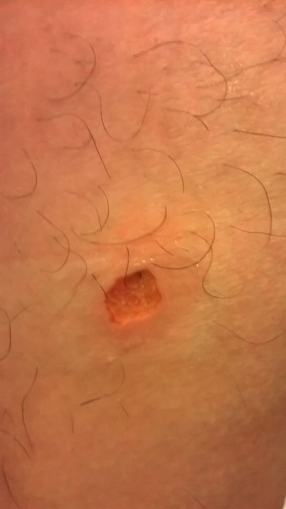
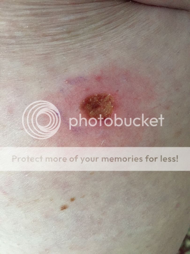
I just got a shave biopsy for a suspicious mole a few days ago and I am waiting to hear the results. I just spotted the mole less than a week ago It was a flat black mole on my thigh and the diamater was just smaller than a pencil eraser. I have not experienced any symptoms such as bleeding, itching, etc. and don't have the risk factors for melanoma. The doctor said it probably was not melanoma in the later stages because of the flatness and he did not see any part of the mole was remaining after but cannot be sure.
My question is if the melanoma already invaded deeper in the skin, shouldn't there be a dark spot on the wound or does it not matter?
Here is a picture of the wound a day after

- Replies
-
-
- July 16, 2015 at 4:41 pm
-
- July 16, 2015 at 4:41 pm
-
- July 17, 2015 at 2:26 pm
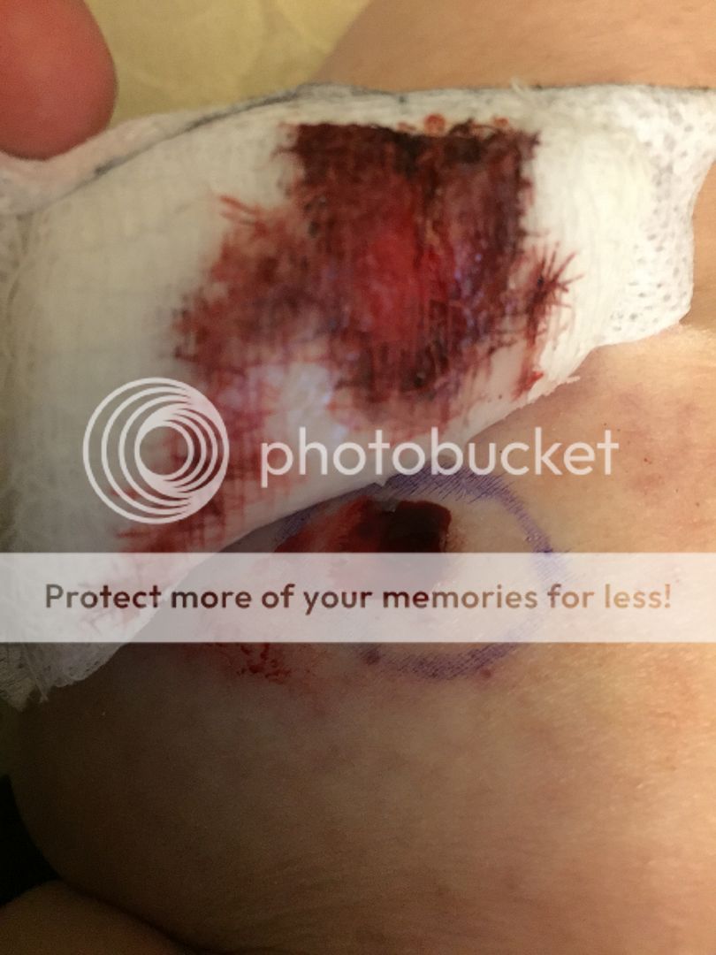
zhanggar, it seems like we're in a similar boat. I had two shave biopsies on Monday for suspicious pigmented lesions, one on my breastbone, one on my lower leg. The dermatologist was concerned about melanoma. I should get the results in a few days. Compared to my biopsy sites, yours looks like its healing well. The site on my breastbone is doing okay, but the one on my lower leg is a bit funky. It bled a lot after the procedure (through the dressing a couple of times) and has subsequently developed a large red area (about 4" diameter) around the biopsy site. The original lesion there was about 1.5cm, flat, and with worrisome characteristics. I've been monitoring the redness–it was definitely expanding, but this morning didn't seem to have changed. I drew a line around it last night so that I could keep an eye on it more easily. My dermatologist said that he really couldn't give an answer, and that pathology was the only way to tell. He did, however, diagnose me with extensive actinic keratoses on my face. I started Imiquimod two days ago, so I'm currently waiting with bated breath for the side effects. So far, after two whole-face applications, my skin feels like its sunburnt and is starting to look patchy and red. Twelve more days to go before I get a break from it.
Anyway, all that to say I send you good wishes for your pathology results.
-
- July 17, 2015 at 8:56 pm
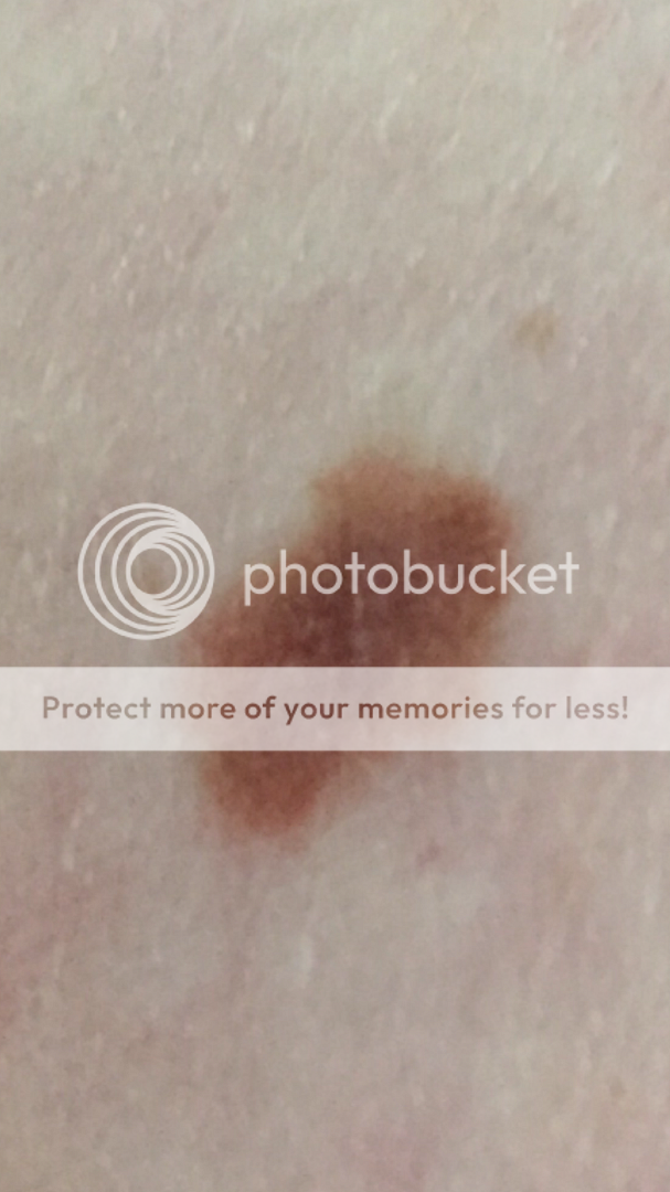
I've had the breastbone lesion for quite a while. In fact, it was first assessed about ten years ago and determined to be unremarkable, but the dermatologist I saw on Monday (for the first time) honed straight in on it as suspicious. That was a smallish lesion, maybe about 7mm, dark. It had started out flat but over the last year or so has developed a raised area. Not nodular, though, as the edges stayed flush with the skin. It also had what appeared to be two much darker areas within it, and on one edge looked as it it was seeping into the surrounding skin. It's gone now, and the biopsy site is doing well.
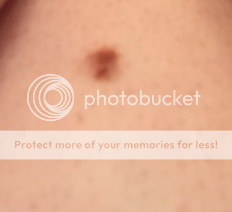
The other lesion was much bigger. I couldn't tell you when I first noticed it, but although it's been there a while now, I haven't always had it, if that makes sense? And, it has most definitely grown. I don't recall specifically, but I'm pretty certain it started life freckle-sized. As of removal day, it was a good 15mm lengthways, 10mm breadthways. Mostly flat with a slight raised area, darker patches of brown, although not as clearly pronounced as on the other lesion, aysmmetrical, and with blurry edges in places.
Neither lesion has ever been itchy or bled.
I have photos of both of them, actually, mostly because I wanted to know what they looked like really close up. I'll post them if you like?
When are you expecting your results? I was told seven business days, which would be around Wednesday of next week.
-
- July 18, 2015 at 3:16 pm
Yea could you post your pictures? I also have a picture of mine although it is not that clear. From what I have read online if the lesion is flat it means it is in the horizontal growth phase and not invaded deeper beneath the skin so that is giving me hope even if it does turn out to be melanoma it will be in the early stage and curable. I only found out about mine about a week ago and fortunately was able to get an appointment to get it removed just 2 days after. I can't remember when it appeared though, I remember checking back of my body a month or two ago and not seeing the lesion but can't be sure because it was located on the back of my thigh and it is hard to look there. I am worried that it may have been there for years and I never noticed it because of the location.
The doctor said I will hear back from them on monday so not much longer now
-
- July 18, 2015 at 3:18 pm
-
- July 18, 2015 at 11:22 pm
Here are my pictures.
This is the one on my leg:
This is the one on my breastbone:
The next three pictures are the biopsy site on my leg the day after, two days after, and three days after (first picture is kind of bloody):
Good luck on Monday!
-
- July 18, 2015 at 11:22 pm
Here are my pictures.
This is the one on my leg:
This is the one on my breastbone:
The next three pictures are the biopsy site on my leg the day after, two days after, and three days after (first picture is kind of bloody):
Good luck on Monday!
-
- July 18, 2015 at 11:22 pm
Here are my pictures.
This is the one on my leg:
This is the one on my breastbone:
The next three pictures are the biopsy site on my leg the day after, two days after, and three days after (first picture is kind of bloody):
Good luck on Monday!
-
- July 18, 2015 at 3:18 pm
-
- July 18, 2015 at 3:18 pm
-
- July 20, 2015 at 11:33 am
Sending positive, good thoughts your way, zhanggar! I'm waiting to call the dermatologists office the second they open…
-
- July 20, 2015 at 7:24 pm
That's great news, zhanggar. Now you can breathe again!
I just managed to speak with the nurse at my dermatologist. The lesion on my calf is a dysplastic nevus, and I'm scheduled for a surgical excision on Friday morning. The lesion on my breastbone is still under review–apparently, the dermatologist sent it for another review, so I've no idea what's going on there. Anyway, I'm relieved that this is just a dysplastic nevus and can be sorted out relatively easily. Phew!
-
- July 20, 2015 at 11:33 am
Sending positive, good thoughts your way, zhanggar! I'm waiting to call the dermatologists office the second they open…
-
- July 20, 2015 at 11:33 am
Sending positive, good thoughts your way, zhanggar! I'm waiting to call the dermatologists office the second they open…
-
- July 18, 2015 at 3:16 pm
Yea could you post your pictures? I also have a picture of mine although it is not that clear. From what I have read online if the lesion is flat it means it is in the horizontal growth phase and not invaded deeper beneath the skin so that is giving me hope even if it does turn out to be melanoma it will be in the early stage and curable. I only found out about mine about a week ago and fortunately was able to get an appointment to get it removed just 2 days after. I can't remember when it appeared though, I remember checking back of my body a month or two ago and not seeing the lesion but can't be sure because it was located on the back of my thigh and it is hard to look there. I am worried that it may have been there for years and I never noticed it because of the location.
The doctor said I will hear back from them on monday so not much longer now
-
- July 17, 2015 at 8:56 pm
I've had the breastbone lesion for quite a while. In fact, it was first assessed about ten years ago and determined to be unremarkable, but the dermatologist I saw on Monday (for the first time) honed straight in on it as suspicious. That was a smallish lesion, maybe about 7mm, dark. It had started out flat but over the last year or so has developed a raised area. Not nodular, though, as the edges stayed flush with the skin. It also had what appeared to be two much darker areas within it, and on one edge looked as it it was seeping into the surrounding skin. It's gone now, and the biopsy site is doing well.
The other lesion was much bigger. I couldn't tell you when I first noticed it, but although it's been there a while now, I haven't always had it, if that makes sense? And, it has most definitely grown. I don't recall specifically, but I'm pretty certain it started life freckle-sized. As of removal day, it was a good 15mm lengthways, 10mm breadthways. Mostly flat with a slight raised area, darker patches of brown, although not as clearly pronounced as on the other lesion, aysmmetrical, and with blurry edges in places.
Neither lesion has ever been itchy or bled.
I have photos of both of them, actually, mostly because I wanted to know what they looked like really close up. I'll post them if you like?
When are you expecting your results? I was told seven business days, which would be around Wednesday of next week.
-
- July 17, 2015 at 9:48 pm
So, since I posted my reply to you I've had a message from the dermatology clinic. Apparently, there is a problem with my path results and I "need further treatment." Of course, I only just got this message, it's Friday early evening, and I can't get hold of anyone to find out what's going on. It's going to be a long weekend.
-
- July 19, 2015 at 11:46 am
Hi Sally
Thinking of you – I also got the dreaded callback ( a voicemail on Thursday last week) – Friday was a public holiday so I still haven't had a chance to talk to the dr. However, this is not my first rodeo – this is my third primary melanoma in 6 months (stage 0, stage 1 and now stage 1). THIS WON'T BE YOU. Very few of us seem to make it into the multiple primary melanoma club.
My advice to you is to do what you have to do to get your situation under control – I am hoping for you that all this means is a wide excision of an early melanoma. A little bit of discomfort, an admirable scar and nothing more. That's my wish for you.
After that, it's important that YOU be just as vigilant as your dr – I had to push for my last two biopsies, guess what, both melanoma. And one was a shave, which is just bad practice because the recommended standard of care (in Australia, for various very good reasons) is a proper 2mm excision.
Here's what can go wrong with a shave: it might go straight through the depth of the melanoma making it nearly impossible to stage. Luckily, that didn't happen to me.
I'm also leary on any kind of partial biopsy e.g., only biopsying part of a larger mole. This is what happened to me: a punch biopsy of the 'scary looking' part of a larger lesion came back as moderately dysplastic (not melanoma) requiring a standard 2mm excision. That 2mm excision came back as a stage 1 melanoma. So for me, any future biopsy is a proper 2mm excision and I would recommend the same to you too. I'll be 'firing' my dr over this incident, already have an appt with a derm booked, because it was psychologically pretty cruel to get a 'relatively all clear' followed by a 'ruh roh my bad stage 1 mel'.
So my second piece of advice to you is: push for excision if you have any doubt whatsoever about a changing lesion (not necessarily ABDCE criteria, just changing) and push for that proper 2mm excision. Shaves and punches are not enough.
Stars
-
- July 19, 2015 at 2:28 pm
Hi stars, thank you so much for your reply. I hope that the way forward for you is as straightforward as can be.
I really appreciate your advice. I originally saw a doctor at University Health Services (I'm a graduate student with student healthcare) at the start of July–we'd been on a family holiday to Florida and while I was putting sun lotion on, I noticed that the lesion on the back of my leg was significantly larger than it had been. Honestly, I wear trousers most of the time and it's in a funny spot towards the back of my calf so although I knew it was there, I hadn't paid attention to it for a long time. The one between my breastbone just looked the same as it always did. Anyway, long stort short, the doctor at UHS said the one on my leg looked suspicious and suggested I see a dermatologist. However, our university hospital (it's a big teaching hospital) only has ONE dermatologist, which is kind of unbelieveable. Without any serious diagnosis, the earliest I could get an appointment with him was early next year, over six months from now! So, doctor suggested I wait until the start of the new semester (end of August) and see another UHS doctor who "knows a bit about skin and can probably do a biopsy." I nodded, booked an appointment, and left.
I knew this wasn't good enough, and decided I was worried enough to call around and find another dermatologist, even though this would take me out of the university health network. I figured it was worh it for the peace of mind. On the second phone call, I got a receptionist who took "suspicious lesion" seriously and squeezed me in to see the dermatologist. This derm is supposed to be good–he has all kinds of certifications and recommendations, so I was pleased to get to see him. I was with him for about ten minutes, fifteen at the most. He diagnosed the actinic keratoses on my face, honed straight in on the two lesions and ordered biopsies. I didn't know enough about biopsies at the time (you can believe I do now) to know that I shouln't have had shave biopsies. I'm hoping that, in the grand scheme of things, this will turn about to be unimportant. Anyway, his nurse did the biopsies, talked me through the side effects of imiquimod, and told me my results would be available online in seven buisness days. Apparently, that's standard procedure for all patient communications. I did ask her what would happen if the pathology showed anything and she said I should call the clinic if that happened–they wouldn't call me. She also suggested that these lesions might just be dysplastic nevi. not necessarily melanoma.
So, off I pootled, and commenced to wait. For some reason that I have yet to fathom, I logged in to the results portal on Friday early evening, and saw that the dermatologist had left a voice mail on Thursday (which didn't come through to my phone, although it should have done) stating that the path results were a problem and I needed further treatment, which would need to be arranged through a nurse. And there we have it. I'm now waiting until the clinic opens tomorrow morning and hoping I can find out exactly what the pathology said, and where to go from here. I'm hoping it's possible that this is a dysplastic nevi and all I need is a couple more mm taking from the biopsy site. I don't know how realistic I'm being with that.
But, I'll be getting back into my university network at the first available opportunity–primarily because I'm not thrilled that a) they did a shave biopsy and b) they've been so lassaiz-faire about communicating with me. Besides, if this anything that needs ongoing attention, I'd rather be with a healthcare network with huge facilities and resources. So, after I get my results tomorrow, I'll be calling the hospital dermatologist to get an appointment in six months, even if all that does is put me in the system for regular skin checks. I'm very glad that something told me to get a biopsy as soon as possible, even if it does still turn out to be nothing. It will have been worth it for the peace of mind. And, if it is something unpleasant, better that I find out now and start treatment than have it lingering for another six months.
Wow. That's quite an essay I wrote. I appreciate your advice and kind words, and am sending positive thoughts back in your direction 🙂
-
- July 19, 2015 at 11:46 am
Hi Sally
Thinking of you – I also got the dreaded callback ( a voicemail on Thursday last week) – Friday was a public holiday so I still haven't had a chance to talk to the dr. However, this is not my first rodeo – this is my third primary melanoma in 6 months (stage 0, stage 1 and now stage 1). THIS WON'T BE YOU. Very few of us seem to make it into the multiple primary melanoma club.
My advice to you is to do what you have to do to get your situation under control – I am hoping for you that all this means is a wide excision of an early melanoma. A little bit of discomfort, an admirable scar and nothing more. That's my wish for you.
After that, it's important that YOU be just as vigilant as your dr – I had to push for my last two biopsies, guess what, both melanoma. And one was a shave, which is just bad practice because the recommended standard of care (in Australia, for various very good reasons) is a proper 2mm excision.
Here's what can go wrong with a shave: it might go straight through the depth of the melanoma making it nearly impossible to stage. Luckily, that didn't happen to me.
I'm also leary on any kind of partial biopsy e.g., only biopsying part of a larger mole. This is what happened to me: a punch biopsy of the 'scary looking' part of a larger lesion came back as moderately dysplastic (not melanoma) requiring a standard 2mm excision. That 2mm excision came back as a stage 1 melanoma. So for me, any future biopsy is a proper 2mm excision and I would recommend the same to you too. I'll be 'firing' my dr over this incident, already have an appt with a derm booked, because it was psychologically pretty cruel to get a 'relatively all clear' followed by a 'ruh roh my bad stage 1 mel'.
So my second piece of advice to you is: push for excision if you have any doubt whatsoever about a changing lesion (not necessarily ABDCE criteria, just changing) and push for that proper 2mm excision. Shaves and punches are not enough.
Stars
-
- July 17, 2015 at 9:48 pm
So, since I posted my reply to you I've had a message from the dermatology clinic. Apparently, there is a problem with my path results and I "need further treatment." Of course, I only just got this message, it's Friday early evening, and I can't get hold of anyone to find out what's going on. It's going to be a long weekend.
-
- July 17, 2015 at 2:26 pm
zhanggar, it seems like we're in a similar boat. I had two shave biopsies on Monday for suspicious pigmented lesions, one on my breastbone, one on my lower leg. The dermatologist was concerned about melanoma. I should get the results in a few days. Compared to my biopsy sites, yours looks like its healing well. The site on my breastbone is doing okay, but the one on my lower leg is a bit funky. It bled a lot after the procedure (through the dressing a couple of times) and has subsequently developed a large red area (about 4" diameter) around the biopsy site. The original lesion there was about 1.5cm, flat, and with worrisome characteristics. I've been monitoring the redness–it was definitely expanding, but this morning didn't seem to have changed. I drew a line around it last night so that I could keep an eye on it more easily. My dermatologist said that he really couldn't give an answer, and that pathology was the only way to tell. He did, however, diagnose me with extensive actinic keratoses on my face. I started Imiquimod two days ago, so I'm currently waiting with bated breath for the side effects. So far, after two whole-face applications, my skin feels like its sunburnt and is starting to look patchy and red. Twelve more days to go before I get a break from it.
Anyway, all that to say I send you good wishes for your pathology results.
-
- July 16, 2015 at 4:41 pm
-
- July 16, 2015 at 5:08 pm
Most melanomas are totally removed via the biopsy. You would not expect to see anything on the skin that would reflect "deeper melanoma" after the biopsy.
-
- July 17, 2015 at 3:20 pm
Most melanomas ARE early stage. The vast majority of melanomas diagnosed are in situ or early stage. I'm not a fan of shave biopsies and will never have one again because they do not get a full skin thickness chunk of skin.
Melanocytes deep in the skin are STAINED on pathology, just so you know. That's so they show up for the pathologist to see. So it would never be likely to see residual melanoma with the naked eye at the depth of a biopsy. Not saying it couldn't happen with an advanced melanoma that was not removed totally, but you are looking at the cellular level when it comes to biopsy tissue.
-
Tagged: cutaneous melanoma
- You must be logged in to reply to this topic.